Kidney Cancer
Kidney cancers can be divided into two groups as the cancers of the part of the kidney that produces the urine (parenchyma) and cancers caused by the collecting duct of the kidney. Here, we will first talk about parenchyma cancers and then collecting duct cancers.
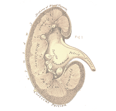
Kidneys are the organs located on the left and right in the back of the abdomen, which filter the blood.
Kidney cancers can be divided into two groups as the cancers of the part of the kidney that produces the urine (parenchyma) and cancers caused by the collecting duct of the kidney. Here, we will first talk about parenchyma cancers and then collecting duct cancers.
Kidney parenchymal cancers make up nearly 3% of the cancers in adults. Male to female ratio is 2:1, and it is mostly common among people aged 50-60. It is known that kidney cancer is more common among people who have certain congenital renal diseases (such as horseshoe kidney and polycystic kidney disease) and certain systemic diseases (such as von Hippel-Lindau syndrome). It is also reported that kidney cancer is more common among people with chronic renal failure. It has been well-proven that smoking increases risk of kidney cancer. In addition, it is reported that overuse of painkillers increases the risk of kidney parenchymal cancer.
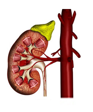
Normal kidney
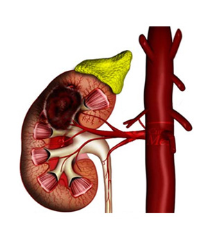
Kidney parenchymal cancer
Diagnosis of Kidney Cancer
Visible blood in urine, pain in the side, and palpable mass, which are the three classic signs in diagnosis of kidney parenchymal cancer, are only present in 10-15% of patients. Most cases are detected randomly during an imaging procedure performed for any other reason.
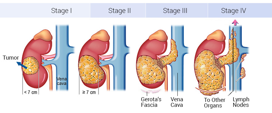
Also, a few patients consult to a physician due to symptoms of metastasis (such as shortness of breath in lung metastasis, bone pain or fractures in bone metastasis), as a result of which, the diagnosis can be established. The rate of kidney cancers randomly diagnosed has been gradually increasing due to widespread use of imaging techniques.
It is reported that 3/4 of kidney cancers are diagnosed randomly today.
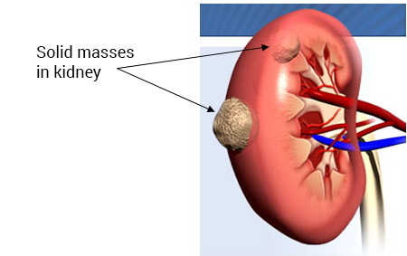
The increase in such rate is associated with the widespread use of ultrasonography in check-up programs. Therefore, there is also increase in the rate of early-diagnosed patients. Detection of a solid mass in the kidney during abdominal ultrasonography suggests presence of kidney parenchymal cancer. Early-diagnosed patients must always be further evaluated with more advanced imaging methods, such as computed tomography (CT) or magnetic resonance imaging (MRI).
Kidney cancers may metastasize and mostly spread to the lungs. Less frequently, it might spread to the liver, bones, adrenal gland, brain and lymph nodes. As the tumor grows in diameter, the risk of metastasis gets higher. If deemed necessary by the physician, further examination should be conducted, such as chest radiography, bone scintigraphy, and positron emission tomography.
Treatment for Kidney Parenchymal Cancers
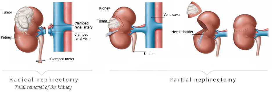
The primary treatment for kidney parenchymal cancers is the surgical removal of the tumor. The most prominent treatment method is removal of the entire kidney together with the surrounding fatty tissue and the adrenal, if necessary, depending on the size and location of the tumor (radical nephrectomy). Performed as traditional open surgeries for years, these surgeries can also be performed with standard laparoscopic or robotic laparoscopic methods today.* Compared to the open surgery, the incision wound is smaller, there is little blood loss, and patients return to their normal lives earlier after the operation in this method. If the diameter of the tumor in the kidney is 4 cm or smaller, it is sufficient to remove only the tumorous area (partial nephrectomy – nephron sparing surgery). Removal of the tumorous area while sparing the normal kidney tissue is possible through not only traditional open surgery but also standard laparoscopic and robotic laparoscopic methods.**
If the the tumor is too large or there is tumor thrombus in the renal vein, traditional open radical nephrectomy should be performed.
Chemotherapy and radiotherapy have very limited use in treatment of kidney parenchymal cancer. New agents are being developed and put in clinical use for this tumor, which is highly resistant to chemotherapy. Radiotherapy, on the other hand, is useful only in treatment of metastatic lesions (bone, brain).
Renal Collecting Duct Cancer: Renal Pelvic Tumor
Collecting duct cancer is a rare form of cancer. Collecting duct cancers, also known as renal pelvic and urothelial tumors, have similar histologic structure with bladder cancers. Smoking and exposure to certain chemicals are known to increase the risk of renal collecting duct cancer.
Most patients notice blood in urine. Sometimes clots may accompany the blood. Pain in the side, nausea and vomiting are among uncommon symptoms. It is highly difficult to diagnose these tumors. Computed tomography (CT) or magnetic resonance imaging (MRI) may be helpful in diagnosing patients with suspected renal collecting duct cancer. However, it may sometimes be necessary to advance through the urethra with a flexible ureteroscope to reach the kidney and take biopsy samples from the tumor. Also, it is possible to treat the tumor with laser if the tumor is small enough.
The ideal treatment for renal collecting duct cancers is the surgical removal of the kidney along with the urethra and the normal tissue around the area where the urethra enters the bladder. Performed with traditional open surgery for years, 2 separate incisions were made in order to remove the kidney and the lower urethra. Today, this surgery can also be performed with laparoscopic method for removal of only the kidney and other tissues, leaving the patient with only an incision wound of 7 cm. Clinical studies show that laparoscopic method can be safely used in this type of cancers. The experience of our staff is also supportive such findings; and 25 patients have been successfully operated on with this method by now.
Since there is a risk of development of a tumor in the bladder of the patients who have undergone a surgery for a collective duct cancer, namely a renal pelvic and urothelial tumor, these patients should be followed up at certain intervals during and after the surgery.
Book an Appointment with Dr. Fatih Atug
Cantact Dr. Fatih Atug
Fatih Atug, M.D.
Urologist and Robotic Surgery Specialist
hidden +90 212 234 5958
hidden +90 532 234 5504
hidden info@fatihatugmd.com
hidden Harbiye Mahallesi, Maçka Caddesi,
Bahriye Apt. No:13 D: 3
Şişli / İSTANBUL
 |
 |
 |


