


Fusion Biopsy
MR-TRUS Fusion Biopsy Technology
Recent scientific studies have shown that prostate cancer detection rate is higher with the fusion biopsy technology than the standard biopsy.
General Information on Prostate Biopsy
Prostate cancer is the second most common cancer in the world and the most frequently diagnosed cancer in Europe and the United States. Diagnosis and screening for prostate cancer is based on serum Prostate-Specific Antigen (PSA) levels, digital rectal examination, and transrectal ultrasound-guided biopsy samples retrieved (TRUS).
During the first years of its application in 1930s, prostate biopsy used to be performed blindly with the help of a finger inserted into the rectum. Introduction of ultrasound-guided prostate biopsy (TRUS) in 1989 was a groundbreaking development, which has led to an increase in the prevalence of the disease.
Standard prostate biopsy (TRUS)
In TRUS guided standard biopsy, tissue samples are collected from the posterior surface of the prostate neighboring the rectum as the ultrasound probe advanced through the rectum provides 2 dimensional images of the prostate (Figure 1). In this technique, samples are retrieved from approximately 12 different zones with mapping method and sent for pathological analysis. Even though the standard biopsy is a method that has been in use for years, the rate of missing prostate cancer is high. With this method, only one in 3-4 patients (30%-40%) is diagnosed with prostate cancer. Patients who cannot be diagnosed and continue experiencing symptoms suggesting prostate cancer are repeatedly exposed to the standard biopsy procedure. Numerous studies, on the other hand, have evidenced the non-negligibly high risk of infection of the biopsy procedure.
Figure 1. Schematic view of standard TRUS biopsy.
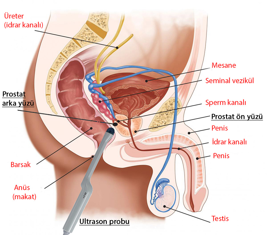
Tissue samples are retrieved from the posterior surface of the prostate leaning against the colon wall with the help of the probe of the ultrasound device advanced through the rectum. Since it is difficult to retrieve samples from the anterior surface of the prostate, the tumors in this zone are mostly missed.
MR-TRUS Fusion Biopsy Technology
Developed recently with the aim of averting the disadvantages of standard biopsies, MR fusion technology provides high rate of cancer detection and allows accurate determination of the stages of the tumors detected.
Fusion biopsy technology is based on combination of multiparametric prostate MR (shortly, mpMR) images with TRUS images. Localized cancer can be detected with high accuracy in the mpMR images of the patients considered to be at risk of prostate cancer. Prior to proceeding with the biopsy procedure, the entire prostate is created in a 3-dimensional format with the help of software, and the localized cancer detected in the prostate tissue can be examined in a 3-dimensional format, as well. The ‘fusion’ procedure is the superimposition of the 3-dimensional MR images on the images received from the ultrasound probe advanced through the rectum (Figures 2 and 3). Thanks to the fusion performed with the help of special software and ultrasound device, it is possible to create a 3-dimensional map of the prostate and retrieve tissue samples from any desired zone in a planned fashion, in contrast to the standard biopsy. With this method, cancer detection rates are higher (approximately 80%) than the standard biopsy.
Figure 2. Artemis™ MR-TRUS fusion biopsy system.

The mpMR images where localized cancers have been marked previously are transferred to the device and superimposed on the ultrasound images to perform the fusion.
Figure 3. Multiparametric MR image of the prostate cancer and Fusion biopsy procedure

The localized cancer detected in the mid-zone of the prostate is shown in images (a), (b) and (c).
(d) – 3-dimensional image of the prostate created by superimposition of the MR image on the ultrasound image (fusion) (The cancerous tissue is shown as the area marked as green. The red lines show the trace of the biopsy needle passing through the cancerous area under the fusion image).
What are the Benefits of Fusion Biopsy compared to Standard Biopsy?
Recent scientific studies have shown that prostate cancer detection rate is higher with the fusion biopsy technology than the standard biopsy. As mentioned above, whereas cancer detection rate is 30-40% in the standard procedure, it reaches 80% in the fusion biopsy. Due to higher accuracy when retrieving biopsy samples, rebiopsies are also avoided. Advancement in the MR technology has also resulted in improvement of the special scoring classification that helps predetermining the clinical significance of the cancer tissue ((PI-RADS scoring (Prostate Imaging-Reporting and Data System)). Thanks to this classification and software developments, the rate of detecting clinically significant prostate cancer –which might result in more deaths and complications- has increased fourfold compared to the standard method.

Book an Appointment with Dr. Fatih Atug
Cantact Dr. Fatih Atug
Fatih Atug, M.D.
Urologist and Robotic Surgery Specialist
hidden +90 212 234 5958
hidden +90 532 234 5504
hidden info@fatihatugmd.com
hidden Harbiye Mahallesi, Maçka Caddesi,
Bahriye Apt. No:13 D: 3
Şişli / İSTANBUL
 |
 |
 |